
Life after craniopharyngioma surgery can bring a lot of mixed feelings. The thought of having a brain tumor can be frightening. For patients and their families, one of the most terrifying situations is not knowing what to anticipate or how life will unfold in the days ahead. It’s the same feeling when one is diagnosed with craniopharyngioma.
It will be crucial for those receiving treatment to understand how to cope with life after craniopharyngioma surgery and how therapy can affect their lives as well as those of their loved ones and caretakers. This article will look at living with craniopharyngioma, symptoms, and surgical options available.
What is Craniopharyngioma?
Craniopharyngioma is an uncommon tumor that develops close to the pituitary gland, a small but powerful organ that controls hormones in the body. This tumor grows slowly and can be difficult to find in its early stages of growth. It is not a cancer, but because of where it develops, it can put pressure on other important brain regions like the optic nerve, pituitary stalk, and hypothalamus.
Craniopharyngioma treatment is necessary if one experiences the condition. The tumor grows close to your pituitary gland, where it can impact structures involved in hormone production and release, as well as many nerves that control eyesight. Craniopharyngiomas can develop into potentially fatal conditions.
Although the tumor itself is not harmful, its effects on the nearby pituitary gland can result in the underproduction of several hormones, which can negatively impact development and other essential body processes.
Craniopharyngioma symptoms
Craniopharyngioma symptoms are associated with disorders that affect the brain, optic nerves, pituitary, and hypothalamus glands. When a craniopharyngioma forms in an adult, the effects on growth and development aren’t noticeable when they are young.
Both adults and children can experience vision problems, especially in areas of their peripheral vision. Healthcare professionals can find it challenging to swiftly determine if the tumor is the origin of these symptoms because they might resemble those of other illnesses.
Diagnosis
A neurological examination is done to properly diagnose symptoms. The neurologist can use the following smell, motor function, swallowing, sensation, balance, and hearing to check if you have craniopharyngioma. If you have vision issues, you will need to perform an eye test, whereas a blood test will be conducted to identify changed hormone levels.
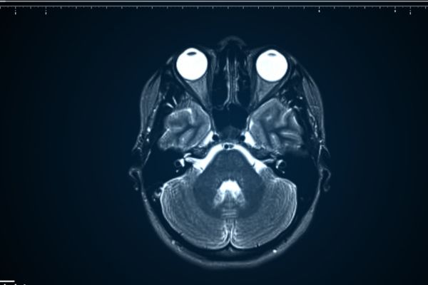
A professional can use imaging tests to ascertain craniopharyngioma location and size. The imaging tests include magnetic resonance imaging (MRI) and computed tomography (CT). When using either test, a contrast enhancement is frequently applied to improve the image’s clarity.
Since most craniopharyngiomas have a calcium buildup, CT scans are very useful for imaging this kind of tumor since they are particularly good at displaying tumors with calcium accumulation.
Craniopharyngioma: When is Surgery Needed?
You need surgery if the tumor results in cognitive impairments, hormonal changes, and elevated intracranial pressure. The initial objective of surgery is for a safe tumor removal and diagnostic confirmation. This would not be feasible, though, if the tumor is near important nearby structures. In that case, the neurosurgeon might remove the tumor partially and suggest radiation therapy following the procedure.
Before deciding how much tumor to remove, neurosurgeons assess each case separately. The strategy and degree of aggressiveness of therapy are determined by a balance between risks and benefits. Significantly, access to neurological care, endocrinologic supervision, and socioeconomic assistance determines the long-term results of surgery in juvenile patients.
Craniopharyngioma: Surgical Options
The transcranial and transnasal methods are the two fundamental surgical techniques used to treat most craniopharyngiomas. A thorough assessment of the tumor’s size and position, the infiltration of structures around it, and the neurosurgeon’s skills all influence the surgical option to choose. The transnasal approach is a less invasive method that accesses the tumor through the natural nasal entry. When the tumor is situated around the midline and base of the brain, it works especially well.
Although less frequently utilized, the more intrusive transcranial approach is beneficial for larger tumors with intricate shapes and locations that call for greater surgical vision and manipulation space. The goal of tumor resection, regardless of approach, is to remove the greatest amount of tumor while maintaining the patient’s functional status.

Life After Craniopharyngioma Surgery
Depending on the nature and outcome of the operation, the recuperation time might last anywhere from a few weeks to several months. Following surgery, individuals may require long-term hormone therapy. Depending on their severity, neurological abnormalities brought on by the tumor or the surgical procedure may negatively impact life following treatment. This can affect social and academic performance, especially in youngsters.
Other uncommon long-term effects of radiation treatment and surgery for craniopharyngiomas may have further neurological ramifications.
Conclusion
Life After Craniopharyngioma Surgery can be difficult, especially if things didn’t go as expected. While some patients can have essentially normal lives, others need long-term medical interventions such as hormone therapy or subsequent therapies because of tumor recurrence.
Managing craniopharyngioma symptoms after surgery will require a supportive medical and caregiver team. To help a patient with a craniopharyngioma, caregivers must have a thorough understanding of their medical needs. While some individuals may need more intense care, others may be able to do simple everyday duties.